In vitro Blood-Brain Barrier Assay Service
The blood-brain barrier (BBB) is formed by endothelial cells of cerebral capillaries and microvessels, coming in close contact with neighboring pericytes and glial cells. As an active interface between the circulation and the central nervous system (CNS), the BBB strictly controls the molecular and cellular traffic between the blood and the brain, thus taking an essential share in providing a steady-state environment for the proper neuronal function.
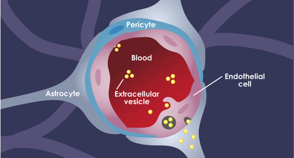 Figure1. The cellular structure of the BBB(Morad et al., 2019)
The BBB protects brain nervous tissue from fluctuating plasma composition from pathogenic agents and maintains homeostasis of the brain parenchyma by restricting non-specific flux of ions, peptides, proteins, and even cells into and out the brain. Many drugs developed to treat CNS disorders are unable to reach the brain parenchyma in therapeutically relevant concentrations. Therefore, it is crucial to characterize the BBB permeability of a drug candidate as early as possible in the developmental drug pipeline. In order to meet this need, in vitro models of the BBB have been developed.
With decades of operational experience and technology platform of BBB research, Creative Bioarray has established multiple in vitro Blood-Brain Barrier Models.
Available BBB Models
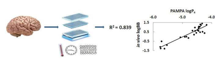 Figure 2. PAMPA(Morad et al., 2019)
The parallel artificial membrane permeability assay (PAMPA) is a non-cell-based, high-throughput permeation model. PAMPA is widely used in the early phase of drug discovery to predict passive diffusion of drug molecules across phospholipid membranes as it is a cost-effective and robust method with good reproducibility(Müller et al., 2015).
Transwell Assays are optimized in PAMPA. By examing effective permeability (Pe) and membrane retention (MR), which detected by LC-MS/MS, to evaluate the permeability of the drug to BBB.
- BBB-MDCK-MDR1 Assay
The human multidrug resistance protein 1 (MDR1)-transfected Madin-Darby canine kidney (MDCK) cell line (MDR1-MDCK) BBB assay can help neuroscience projects identify MDR1 efflux liability in the early discovery stage to avoid MDR1-related brain impairment issues(Di et al., 2013). Although the MDCK-BBB model is not based on brain endothelial cells, it still has an excellent ability to detect drug penetration.
Transwell Assays are optimized in MDCK-MDR1 assay. By examing percent recovery, apparent permeability coefficient (Papp), and efflux ratio, which detected by LC-MS/MS, to evaluate the drug penetration to BBB.
NOTICE: MDCK cell line mentioned above has two subtypes(Feng et al., 2019).
| MDCK I | Derived from low passage parental cell line (NBL-2). Type I cells display high TER values (> 1000 Ω•cm2), are negative for the tight junction claudin-2, and are reported to display adrenalin, vasopressin, and prostaglandin E sensitive adenyl cyclase. |
| MKCD II | Isolated from high passage parental cell line (NBL-2). Type II cells display low TER values (~100 Ω•cm2) and are positive for claudin-2. They are larger and taller than MDCK I cells and lack gap junctions unless modified. |
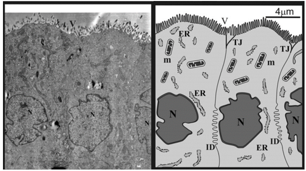 Figure 3. Electron micrographs of rat MDCK-MDR1 cell cytoarchitecture. ER, endoplasmatic reticulum; ID, interdigitations; m, mitochondrion; N, nucleus; TJ, tight intercellular junctions; V, microvilli(Hellinger et al., 2012).
- Co-culture Systems
Creative Bioarray has established in vitro Blood-Brain Barrier Model using primary brain endothelial cells, subjected to in vitro Blood-Brain Barrier assays, such as oxygen-glucose deprivation to mimic stroke conditions.
Astrocytes can induce the formation of interendothelial TJs, a fundamental characteristic of the BBB. A significant number of currently used in vitro BBB model is composed of co-culture of brain endothelial cells with astrocytes.
We can use both double and triple co-culture systems for in vitro BBB model generation based on the culture of endothelial cells with astrocytes and pericytes.
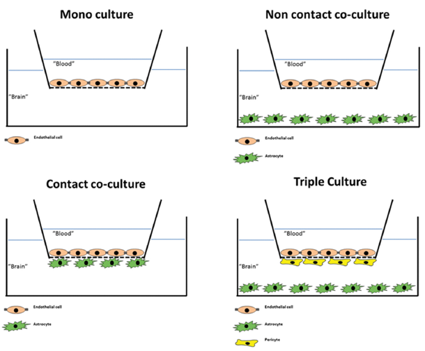 Figure 4. Commonly used configurations used for culture of brain endothelial cells(Helms et al., 2016).
Quotation and ordering
If you have any special needs in our In Vitro Blood-Brain Barrier Assay Service, please contact us for this particular service. We also provide in vivo BBB assays using mice and rats.We also provide. Let us know what you need, and we will accommodate you.
We look forward to working with you in the future.
References
- Di, L.; et al. Demystifying Brain Penetration in Central Nervous System Drug Discovery. Journal of Medicinal Chemistry, (2013), 56(1), 2-12.
- Feng, B.; et al. Validation of Human MDR1-MDCK and BCRP-MDCK Cell Lines to Improve the Prediction of Brain Penetration. Journal of Pharmaceutical Sciences, (2019),108(7), 2476-2483.
- Hellinger, É.; et al. Comparison of brain capillary endothelial cell-based and epithelial (MDCK-MDR1, Caco-2, and VB-Caco-2) cell-based surrogate blood–brain barrier penetration models. European Journal of Pharmaceutics and Biopharmaceutics, (2012),82(2), 340-351.
- Helms, H. C.; et al. In vitro models of the blood–brain barrier: An overview of commonly used brain endothelial cell culture models and guidelines for their use. Journal of Cerebral Blood Flow & Metabolism, (2016), 36(5), 862-890.
- Morad, G.; et al. Tumor-Derived Extracellular Vesicles Breach the Intact Blood-Brain Barrier via Transcytosis. ACS Nano, (2019), 13(12), 13853-13865.
- Müller, J., et al. Tuning the predictive capacity of the PAMPA-BBB model. European Journal of Pharmaceutical Sciences, (2015), 79, 53-60.
