Featured Products
Our Promise to You
Guaranteed product quality, expert customer support

JVM-3
- Specification
- Recommended Products
Immunology: CD3 -, CD10 -, CD13 -, CD19 +, CD20 +, CD34 -, CD37 +, cyCD79a +, CD80 +, HLA-DR +, sm/cyIgG
- Background
- Scientific Data
- Q & A
- Customer Review
- Product Information Sheet
JVM-3 cells are a cell line derived from the peripheral blood of a 73-year-old man diagnosed with B cell prolymphocytic leukemia (B-PLL) at the time of diagnosis. The cell line was established through Epstein-Barr virus (EBV) transformation during treatment with the phorbol ester TPA (12-O-tetradecanoylphorbol-13-acetate). JVM-3 cells have been extensively studied and utilized as a model for investigating B-cell leukemia and related oncogenic pathways.
Establishing JVM-3 cells has provided researchers with a valuable tool for studying the molecular mechanisms underlying B-PLL and the role of BCL2 and BCL3 in leukemogenesis. Researchers use JVM-3 cells to investigate the impact of these proto-oncogenes on cellular signaling pathways, apoptosis resistance, and potential therapeutic strategies targeting BCL2 or BCL3.
Furthermore, the EBV transformation of JVM-3 cells during TPA treatment allows researchers to explore the influence of viral infection and TPA-induced cellular changes on leukemic transformation and disease progression. JVM-3 cells provide a platform for studying the interplay between viral factors, oncogenic signaling, and leukemia development.
Functional Characterization of SIRT1 and SIRT2 in B-Cell Chronic Lymphocytic Leukemia (CLL)
Sirtuins (SIRT) proteins play an important role in the survival and drug resistance of cancer cells. SIRT1 and SIRT2 protein expression were quantified in fresh CLL samples from 9 patients and 2 cell lines derived from B-PLL (JVM-3 and MEC-2) using Western blotting. As shown in Fig. 1A, both SIRT1 and SIRT2 proteins were overexpressed in all the CLL samples examined, however, SIRT2 levels were lower than SIRT1 in most samples (Fig. 1B). By contrast, there was little difference in the protein levels of SIRT1 and SIRT2 in the 2 PLL cell lines JVM-3 and MEC-2 as measured by Western blotting (Fig. 1C) followed by quantitation of relative protein expression (Fig. 1D). These results demonstrate that both SIRT1 and SIRT 2 are overexpressed in CLL patients and PLL cell lines.
The effect of SIRT inhibitors, sirtinol, and EX-527 in PBMC was tested by analyzing cell viability by WST1. EX-527 and sirtinol treatment inhibited cell growth in all the samples tested with an IC50 ranging from 50-100 µM for Ex-527 and 10-20 µM for sirtinol. Cell viability was significantly inhibited in the presence of both inhibitors in a dose-dependent manner at 24-72 hr. (Fig. 2A). Similar effects of EX-527 and sirtinol were observed on JVM-3 and MEC-2 cell lines (Fig. 2B). Both drugs induced significant apoptosis at all the concentrations tested in most of the samples (Fig. 2C) as well as in the cell lines (Fig. 2D). Taken together, these data suggest that inhibition of SIRT1 results in cellular apoptosis in CLL patient samples and relevant cell lines.
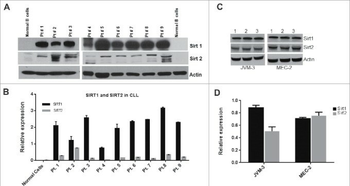 Fig. 1 SIRT1 and SIRT2 are overexpressed in CLL cell lines (JVM-3 and MEC-2). (Bhalla S and Gordon LI, 2016)
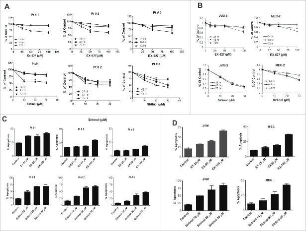 Fig. 2 Pharmacological inhibition of Sirtuins affects cell viability and induces apoptosis in CLL cell lines (JVM-3 and MEC-2). (Bhalla S and Gordon LI, 2016)
Ibrutinib synergizes with MDM-2 inhibitors in promoting cytotoxicity in B-CLL
The potential efficacy of the Ibrutinib/Nutlin-3 combination was evaluated in vitro in a panel of B leukemic cell lines (EHEB, JVM-2, JVM-3, MEC-1, MEC-2) and primary B-chronic lymphocytic leukemia (B-CLL) patient samples. As shown in Fig. 3, the combined treatment induced a reduction of cell viability significantly higher for the single drugs in all B leukemic cell lines analyzed. The induction of apoptosis, rather than the cell cycle block mainly accounted for the synergistic anti-leukemic activity of the Ibrutinib/Nutlin-3 combination. The analysis of apoptosis showed that Ibrutinib alone induced modest levels of apoptosis in all cell lines, while the Ibrutinib/Nutlin-3 combination significantly increased the percentage of apoptosis for the treatment with the single drugs used alone, in both p53wild-type and p53mutated B leukemic cell lines (Fig. 4).
It is well known that the pathogenesis of B-CLL is characterized by alterations of cellular signaling pathways. Exposure of B leukemic cell cultures to Ibrutinib resulted in inhibition of BTK auto-phosphorylation at tyrosine 223 (caused by the inhibition of kinase activity by the binding of Ibrutinib) coupled to a significant reduction of the phosphorylation of the MAPK survival pathway modulators ERK1/2, starting at early time points post drug exposure (Fig. 5A). Moreover, by assessing phosphorylation levels through magnetic-plex assays, a rapid down-modulation of the PI3K survival pathway was observed, as documented by the reduction of both Akt and m-TOR phosphorylation, which was validated also by Western blotting (Fig. 5B). No significant modulation of the p53 pathway was observed in p53mutated B cells following any treatment (Fig. 5C). Western blotting analysis on cellular lysates from B-leukemic cell lines highlighted that the exposure to Ibrutinib was associated to up-regulation of phospho-H2A.X, responsible for the DDR-signal amplification, and that this response was further enhanced by the combination with Nutlin-3 (Fig. 5D).
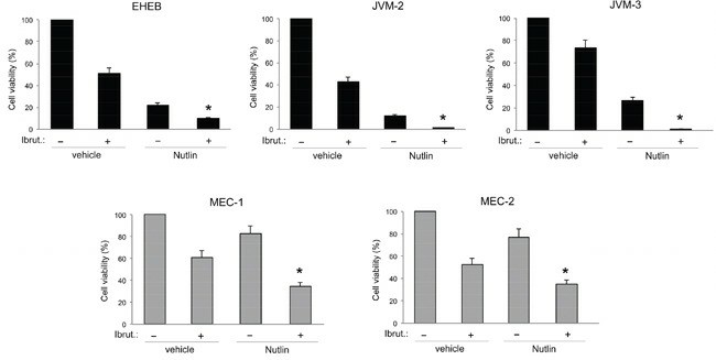 Fig. 3 Evaluation of cytotoxic effect in response to Ibrutinib and Nutlin-3 used alone or in combination in B leukemic cell lines. (Voltan R, et al., 2016)
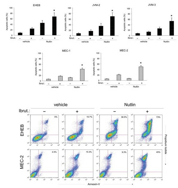 Fig. 4 Effects of Ibrutinib/Nutlin-3 combination on cell apoptosis in B leukemic cell lines. (Voltan R, et al., 2016)
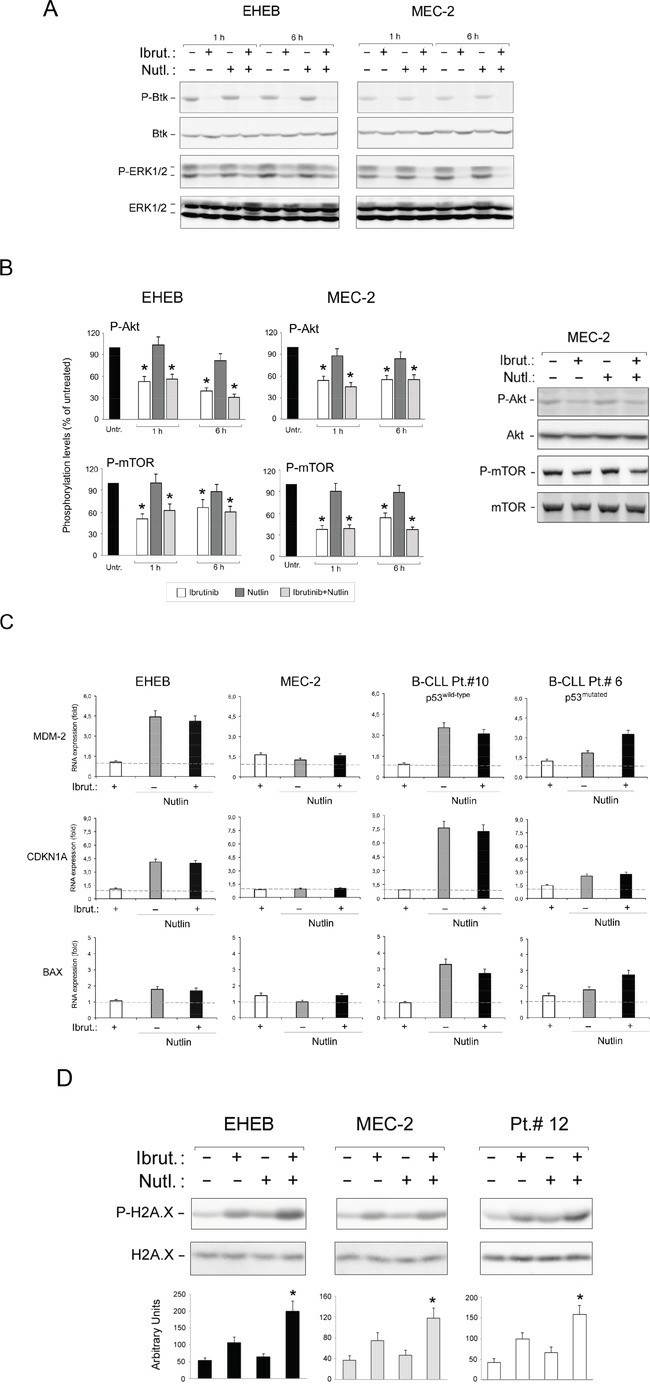 Fig. 5 Intracellular pathway mediating the anti-leukemic activity of Ibrutinib/Nutlin-3 combination. (Voltan R, et al., 2016)
A cell viability assay measures the number (relative or absolute) of living cells in a sample. Viability is a function of cellular proliferation and death. Cytotoxicity assays measure the number (relative or absolute) of dead cells.
JVM-3 cells were established through EBV (Epstein-Barr virus) transformation during treatment with the phorbol ester TPA (12-O-tetradecanoylphorbol-13-acetate).
JVM-3 cells express mRNA for the proto-oncogenes BCL2 and BCL3. These genes are involved in several cellular processes, including apoptosis regulation, cell cycle control, and the immune response.
Studying JVM-3 cells provides insights into the molecular mechanisms underlying B-prolymphocytic leukemia and the role of BCL2 and BCL3 in leukemogenesis. These cells serve as a valuable model for investigating oncogenic signaling pathways and potential therapeutic strategies.
Average Rating: 5.0 | 3 Scientist has reviewed this product
Top-notch quality
The durability of cell products in various conditions has greatly assisted in my research. Kudos to the provider's services.
22 Oct 2022
Ease of use
After sales services
Value for money
Helpful documentation
The provided documentation, including growth protocols and quality control data, was comprehensive and helpful.
21 Sep 2023
Ease of use
After sales services
Value for money
Excellent services
The customer service was excellent, promptly addressing any inquiries and providing support throughout the ordering process. I highly recommend Creative Bioarray for its reliable JVM-3 cell products.
13 Jan 2024
Ease of use
After sales services
Value for money
Customer Support & Price Inquiry
