Isolation and Characterization of Exosomes from Non-Small Lung Cancer Cells
Journal of Cancer Research and Clinical Oncology. 2023 Aug; 149 (10): 7505-7514.
Authors: Mahgoub EO, Abdella GM.
INTRODUCTION
Exosomes are nanovesicles that are constantly secreted by cells into the human body's circulation. Their described sizes vary considerably, which mirrors the difference in the isolation techniques. In this study, we have identified improved methods for isolating exosomes from non-small lung cancer cells (NCI1975). Generally, isolated exosomes are characterized by their morphology, size, surface marker protein expression, purity, and yield concentration.
METHODS
- Cell culture. NCI1975 cells were grown to 70% confluence, and then the cells were washed twice with PBS and 14 ml of DMEM with no serum. NCI1975 cells were incubated for 24 h, and after 48 h, 14 ml of conditioned medium was harvested from 70% confluent NCI1975 cells. The conditioned medium was centrifuged at 4000×g for 10 min to remove detached cells.
- Exosome isolation. Additionally, 4 ml of collected supernatant was centrifuged at 53,000 g rpm at 4°C for 2 h. The supernatant was carefully removed, and crude exosome-containing pellets were resuspended in 1 mL of ice-cold PBS and pooled. The second round of ultracentrifugation at 53,000 rpm at 4°C for 90 min was carried out, and the resulting exosome pellet was resuspended in 50 µL of PBS or loading buffer before use in microscopes. The exosomes collected after multiple rounds of ultracentrifugation were then stored in PBS buffer as starting material and subjected to ultrafiltration.
- Chemical characterization of exosome. Characterization of extracted exosomes using western blot analysis, and protein fraction size was detected using 10% SDS-PAGE.
- Morphological characterization of exosome. The morphology was analyzed by transmission electron microscopy (TEM) and scanning electron microscopy (SEM). TEM is a morphological characterization method used to characterize exosomes. Scanning microscopy samples were prepared by preparing helium ion slides from exosome samples.
- Browse our recommendations
Creative Bioarray provides professional products and services, including but not limited to the following.
| Product/Service Types | Description |
| Exosome Isolation and Purification | Creative Bioarray provides reliable techniques for exosome isolation from different sample matrices. |
| Exosome Identification | Our expertise characterizes isolated exosomes in many different ways including particle size, quantity, specific antibodies, and more. |
| Transmission Electron Microscopy (TEM) Service | Creative Bioarray has scientists and imaging laboratories to perform the full comprehensive Transmission Electron Microscopy (TEM) and Scanning Transmission Electron Microscopy (STEM) services for the biological sciences and clinical research. |
RESULTS
- In this study, we investigated the purity and stability of exosome proteins after excessive isolation methods. Exosomes were isolated using ultracentrifugation followed by ultrafiltration, and then special preservation techniques improved exosome purity and yield concentration. The products of ultracentrifugation techniques alone and ultrafiltration alone, compared with the combination of the three methods, were incredibly different in purity and concentration. Three conventional exosome markers (e.g., CD9, CD81, and CD63) were analyzed to characterize the expression of the exosome's significant proteins.
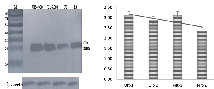 Fig. 1 Band of 28 kDa of exosomes has been treated with CD9 of rabbit anti-mouse.
- The morphological characterization results showed that the exosomes were small vesicles. Moreover, extracellular vesicles isolated from normal cells showed a round morphology consistent with exosome sizes and shapes isolated from normal cells. In addition, the morphological shape of exosomes was computed to vary in size, ranging from 30 to 200 nm in diameter. In contrast to exosomes, apoptotic vesicles and necrotic bodies isolated from NSCLC (NCI 1975) cells displayed irregular shapes and heterogeneous size distributions.
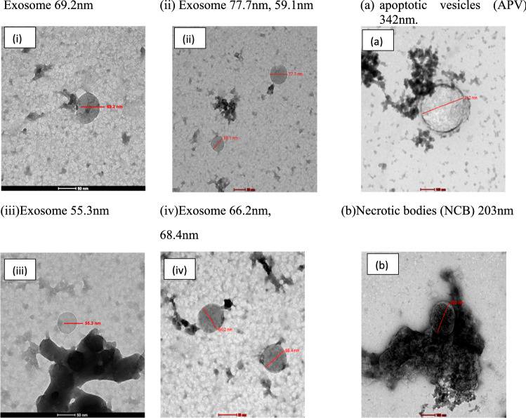 Fig. 2 TEM was used for morphology characterization of the exosome.
SUMMARY
This study has demonstrated improved methods for isolating exosomes from non-small lung cancer cells, which address the problems characterized by exosome morphological and chemical methods. The isolated exosomes were characterized using TEM and SEM was added for further assurance of the investigation results.
RELATED PRODUCTS & SERVICES

Reference
- Mahgoub EO, Abdella GM. (2023). "Improved exosome isolation methods from non-small lung cancer cells (NC1975) and their characterization using morphological and surface protein biomarker methods." J Cancer Res Clin Oncol. 149 (10), 7505-7514.
