Establishment of a New Pancreatic Cancer Cell Line
World Journal of Gastroenterology. 2023 May 7; 29 (17): 2642-2656.
Authors: Xu H, Chai CP, Miao X, Tang H, Hu JJ, Zhang H, Zhou WC.
INTRODUCTION
- Ampullary carcinoma is a relatively rare tumor, accounting for 0.2% of gastrointestinal cancers but 20% of all periampullary cancers. Recently, the incidence rate of ampullary cancer has gradually increased.
- So, establishing an ampullary carcinoma cell line, conducting in-depth research on the pathogenesis and biological characteristics of ampullary carcinoma, and then investigating therapeutic targets to formulate effective treatment strategies for patients with ampullary carcinoma are currently required.
METHODS
- The tissue samples were collected from a patient with ampullary cancer. In this study, experiments were conducted on 4-6-week-old BALB/c nude female mice, weighing 16-20 g.
- Establishment of the cell line. The small woven pieces were mixed with type II collagenase (Gibco) and neutral protease (Invitrogen) and digested in a shaking table at 37°C. The sample was centrifugated at 300 × g for 3 min and the supernatant was discarded. The precipitate was added to a complete medium [RPMI-1640 + 10% fetal bovine serum (FBS) + 1% penicillin-streptomycin] and uniformly inoculated on a six-well plate (NEST).
- Cell growth curve. The cell density of DPC-X1 cells in the logarithmic growth phase (P20) was adjusted to 1 × 10⁴/mL after trypsin digestion. Inoculate 0.1 mL of sample into the reagents of the 96-well plate cell counting kit. The cell growth curve was plotted with time as the horizontal axis and absorbance value as the vertical axis.
- Analysis of short tandem repeats. DPC-X1 cells together with their primary tumor tissue were subjected to short tandem repeat sequence (STR) analysis to determine the correlation between the cells and the primary tumor tissue.
- Karyotype analysis. DPC-X1 cells in the logarithmic growth phase (P40) were treated with 0.25 μg/mL colchicine for 6 h overnight at 37°C. The mitotic phase cells with good dispersion and moderate staining were selected for karyotype analysis.
- Transmission electron microscopy (TEM). DPC-X1 cells in the logarithmic growth phase (P45) were digested, centrifuged, fixed with an electron microscope fixative, and stored and transported at 4°C. Finally, the images were observed under the TEM.
- Immunohistochemical staining. The paraffin sections of primary tumors, transplanted tumors, and organ-like tissues were prepared and incubated at 60°C overnight. Subsequently, 3% hydrogen peroxide solution was incubated at 37°C for 15 min to block the activity of peroxidase; 100 μL of normal goat serum was dripped and then kept sealed at 37°C for 15 min. Dehydration and xylene transparency were carried out after hematoxylin counterstaining, and the samples were observed under the microscope after neutral resin sealing.
- Browse our recommendations
As an established leader in the field of cell science, Creative Bioarray offers a comprehensive range of services related to cell line establishment for our client's research, such as Cell Line Authentication, Karyotyping (G-Banded) Service, Transmission Electron Microscopy (TEM) Service, Immunohistochemistry (IHC), Immunofluorescence (IF) Service, Special Staining Services, and others.
RESULTS
- Through primary culture and subculture of primary tumor tissue of ampullary carcinoma, an ampullary carcinoma cell line, named DPC-X1, was successfully established. The cell morphology and growth mode remained unaltered even though it passed to the 80th generation.
- DPC-X1 cells proliferated vigorously and grew stably in RPMI-1640 medium with 10% FBS. The DNA typing results demonstrated that DPC-X1 is a new human ampullary carcinoma cell line with the same origin as that of the primary tumor tissue. Karyotype analysis showed that DPC-X1 cells were mainly sub-tetraploid, with large differences in chromosome number and morphology.
- Under the TEM, the DPC-X1 nucleus appeared large and deformed, and its number was increased. DPC-X1 was sensitive to oxaliplatin and paclitaxel and resistant to fluorouracil and gemcitabine. DPC-X1 was able to rapidly form xenograft tumors under the skin of nude mice, and the tumor formation rate was 100%. Hematoxylin-eosin staining showed visible multinucleated cells and megakaryocytes, exhibiting typical features of malignancy.
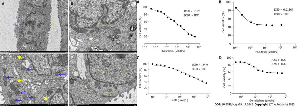 Fig. 1 Left: Ultrastructure of DPC-X1 under a transmission electron microscope; Right: Drug sensitivity test.
SUMMARY
In summary, the study reports a novel human mixed-type ampullary carcinoma cell line DPC-X1 developed from a patient's tumor. This cell line provides a new experimental model for studying the biological and molecular mechanisms of ampullary carcinoma and developing new therapeutic drugs.
RELATED PRODUCTS & SERVICES

Reference
- Oreff GL, et al. (2023). "Immortalized murine tenocyte cells: a novel and innovative tool for tendon research." Sci Rep. 13 (1), 1566.
