Deep Immunohistochemistry for 3D Histology
Cell Reports Methods. 2023 Apr 20; 3 (5): 100458.
Authors: Yau CN, Lai HM, Lee K, Kwok AJ, Huang J, Ko H.
INTRODUCTION
Deep immunohistochemistry (IHC) is a nascent field in three-dimensional (3D) histology that seeks to achieve thorough, homogeneous, and specific staining of intact tissues for visualization of microscopic architectures and molecular compositions at large spatial scales. Despite the tremendous potential of deep IHC in revealing molecule-structure-function relationships in biology and establishing diagnostic and prognostic features for pathological samples in clinical practice, the complexities and variations in methodologies may hinder its use by interested users.
Technical Approaches
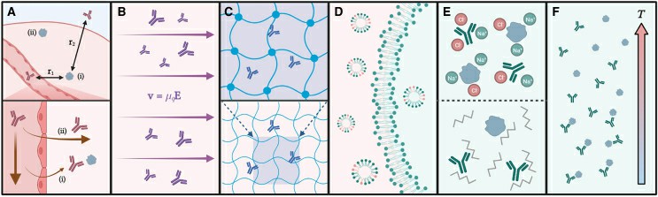
Fig. 1 A graphical summary of deep immunostaining technical approaches.
- A pipeline for deep immunostaining typically consists of several modules: (1) sample fixation, (2) optional tissue pre-treatment (e.g., hydrogel embedding, dehydration), (3) permeabilization, and (4) immunostaining. In practice, the majority of deep IHC methods are coupled with optical clearing, which typically adds an extra module of (5) refractive index matching, followed by (6) volumetric imaging, although this is not strictly necessary when tomography by serial sectioning is employed. The variety of deep IHC approaches stems from the combination and modification of each of these modules.
- Existing deep immunolabeling approaches can be broadly classified into two categories based on their main principle adopted to enhance Ab penetration: (1) modulation of Ab-Ag binding, and (2) facilitation of Ab diffusion and/or advective transport. The shared principle behind techniques based on modulation of Ab-Ag interaction is that by temporarily inhibiting Ab-Ag binding, less Ab depletion occurs, and more mobile, Abfs are available to diffuse deeper into tissue. This has been accomplished in different ways, using detergents (e.g., SDS, NaDC), non-specific denaturants (e.g., urea, Quadrol), tuning pH, heating, or combinations of these. On the other hand, Ab movement can be enhanced by tissue permeabilization, reducing the distance to reach Ag targets, and/or use of externally applied force fields to create advective transport.
Quality Control
For fair and quantitative evaluations of existing and newly developed methods, three metrics are of particular interest: penetration depth of probes, staining homogeneity, and staining specificity.
- Penetration depth of probes
A sufficient penetration depth is a prerequisite for examining the homogeneity and specificity of staining in large tissues. Upon staining, signal intensities of voxels with labeled markers increase, and a subset of them exceeds the detection threshold of the optical system used. Depending on the experimental objective, suprathreshold voxels may be grouped as units (e.g., segmented as cells). The penetration depths of probes can then be defined as the maximal value of the shortest distances between the suprathreshold voxel units and the tissue surface. - Staining homogeneity
Quantitative 3D IHC for many tissue markers necessitates homogeneous staining. Staining homogeneity is determined by the extent of equal Ab accessibility of Ags across the tissue. One or more tissue markers with uniform distribution across depths should be chosen for IHC, and a high staining homogeneity is demonstrated when maximal signal homogeneity (or minimal signal variability) versus depth is obtained. Quantification can be achieved by examining marker-associated voxel signal intensity versus distance from the nearest tissue surface, followed by appropriate curve-fitting and parameter extraction. - Staining specificity
Many Ags have heterogeneous spatial distributions, and authentic results of immunolabeling should faithfully reveal their characteristic patterns in tissues. Staining specificity against a given Ag or marker can thus be defined as the resemblance of the staining pattern obtained via deep IHC methods to the ground-truth distribution in thin sections. The experimental approach and quantifications required to obtain such information, along with penetration depth and staining homogeneity, are outlined in the next section.
Creative Bioarray Relevant Recommendations
Creative Bioarray offers a comprehensive IHC service from project design, and marker selection to image completion and data analysis. We are dedicated to satisfying every customer and assisting them to achieve their specific research goals.
RELATED PRODUCTS & SERVICES
Reference
- Yau CN, et al. (2023). "Principles of deep immunohistochemistry for 3D histology." Cell Rep Methods. 3 (5), 100458.
