A Novel Hilar Cholangiocarcinoma Cell Line, CBC3T-1
Human Cell. 2024 Jan; 37(1): 364-375.
Authors: Bai M, Jiang N, Fu W, Huang C, Tian L, Mi N, Gao L, Ma H, Lu Y, Cao J, Zhang C, Yue P, Zhang Y, Lin Y, Meng W, Li X.
INTRODUCTION
Cholangiocarcinoma (CCA) is a group of malignant heterogeneous cancers arising from the biliary tree. The tumor is characterized by insidious onset, a high degree of malignancy, a poor prognosis, and a high recurrence rate. Immortalized cancer cell lines are the best and easiest models for in vitro cancer research.
METHODS
- The donor patient was a 71-year-old male with hCCA. The patient had elevated tumor marker levels, and computed tomography (CT) images showed circumferential thickening and mild enhancement of the perihilar bile duct with luminal narrowing. A diagnosis of hCCA was considered. No preoperative radiotherapy or chemotherapy was administered, and the patient underwent resection of the hCCA. The tumor specimen showed a circumferential soft-tissue perihilar mass. The postoperative pathological diagnosis of this patient's tumor tissue was moderately differentiated adenocarcinoma of the bile duct.
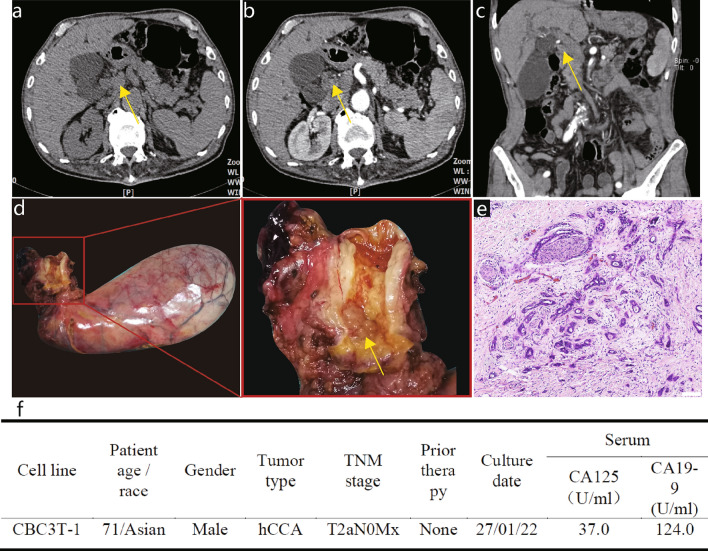 Fig. 1 Clinical and pathological profile of CBC3T-1.
- The cells were cultured in RPMI 1640 medium containing 10% FBS, 100-U/mL penicillin, and 100-mg/mL streptomycin, and placed in an incubator with a constant temperature of 37°C and 5% CO2. For the detection of cellular mycoplasma, the medium of CBC3T-1 cells was collected and assayed according to the mycoplasma detection kit.
- Cells were incubated for 2 h using 0.4 µg/ml colchicine. Then cells were collected and incubated with 0.075 M KCl for 30 min (37°C) and fixed 3 times with methanol: acetic acid (3:1) at room temperature for 10 min. Slides were then prepared and stained with Giemsa. Representative images of chromosome sets were obtained for karyotype analysis.
- Cells were digested and cultured in 96-well plates until they were attached to the surface. Live cell imaging was performed using an imaging system under a 4 × objective. Label-free live cell proliferation measurements were achieved by capturing two high-contrast brightfield images at each 2-h time point.
- Browse our recommendations
| Product/Service Types | Description |
| Cell Line Authentication | Creative Bioarray provides a convenient STR-based method for cell line identity confirmation and for the detection of intra-species contamination in cultured cell lines. |
| Cell Line Testing and Assays | Creative Bioarray offers a wide range of cell line testing and assays from cell viability and proliferation to cellular phosphorylation assays. |
| Tumor Cells | Creative Bioarray's tumor cell panels are based on key components of cell signaling pathways or cancer genes. |
| Tumor Cells Media | With our expertise and dedication to advancing cancer research, we specialize in developing and manufacturing specialized media formulations optimized for the growth and maintenance of tumor cells. |
RESULTS
- A cell line from the primary tumor tissue of a 71-year-old male patient with hCCA was established and designated it CBC3T-1. To date, CBC3T-1 cells have been maintained in single-molecule culture for over 60 generations. We performed STR analysis of 21 loci to determine the identity of the cell line and to exclude cross-contamination. The genomic identity of the CBC3T-1 cell line was also confirmed by comparison with the genetic profile of the primary tumor. These results suggest that CBC3T-1 is a novel hCCA cell line.
- CBC3T-1 cells were irregularly polygonal, with one or more oval nuclei and visible nucleoli. When grown at high density, the cells accumulated and showed loss of contact inhibition, indicating cellular malignancy. The doubling time of CBC3T-1 cells was 52 h. Meanwhile, we performed live cell imaging of CBC3T-1 cells to dynamically observe cell proliferation. CBC3T-1 cells were not contaminated with mycoplasma. Karyotype analysis of a representative single cell from CBC3T-1 shows a hyperploid cell line with a karyotype abnormality and a chromosome number between 69 and 73.
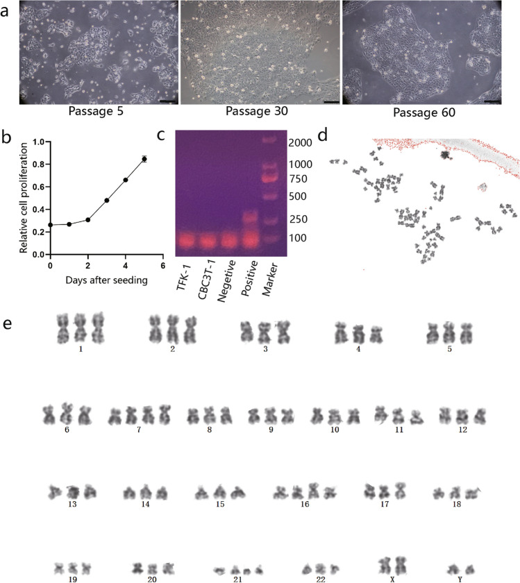 Fig. 2 Characterization of CBC3T-1.
- The ability of established CBC3T-1 cells to form spheres was assessed, with cells forming spheres in a low-adherent plate. The spheroid formation assay can be used to preliminarily judge the tumorigenicity of cells in vitro. To further characterize the tumorigenic properties of the CBC3T-1 cell line, TFK-1 cells were used as controls for further analysis. Wound-healing assay results showed higher levels of wound repair by CBC3T-1 cells at 12 h or 24 h. In addition, CBC3T-1 cells had a stronger migratory and invasive capacity than TFK-1 cells. Finally, we performed colony formation assays and found that CBC3T-1 cells exhibited enhanced clonogenic capacity.
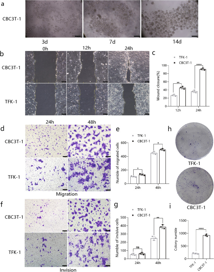 Fig. 3 Characterization of CBC3T-1 cell behavior.
SUMMARY
A naturally immortalized hCCA cell line, CBC3T-1, was established. The CBC3T-1 cell line was cultured for over 60 passages. Thorough analysis showed that CBC3T-1 cells share characteristics similar to original tumor cells from patients with cholangiocarcinoma and display a stable phenotype, including features of epithelial origin, stem cell-like properties, as well as a high invasive and migratory capability and tumorigenicity in mice.
RELATED PRODUCTS & SERVICES

Reference
- Bai M, et al. (2024). "Establishment and characterization of a novel hilar cholangiocarcinoma cell line, CBC3T-1." Hum Cell. 337 (1): 364-375.
