Chick Chorioallantoic Membrane (CAM) Angiogenesis Assay
The chick embryo chorioallantoic membrane (CAM) is an extraembryonic membrane that acts as a gas exchange surface and its function is supported by a dense capillary network. Due to its extensive vascularization and easy accessibility, the CAM has been broadly used to study the morphological function of the angiogenesis process in vivo and to investigate the efficacy and mechanisms of pro-angiogenic and anti-angiogenic molecules.
The CAM angiogenesis assay is performed by implanting a membrane or coverslip containing the compound of interest on the chick embryo chorioallantoic membrane through a hole cut in the egg shell. Subsequently, the CAM is fixed and the blood vessels are quantified by counting the number of blood vessel branch points. The main advantages of CAM model are high vascularization properties, high reproducibility, simplicity, and cost effectiveness.
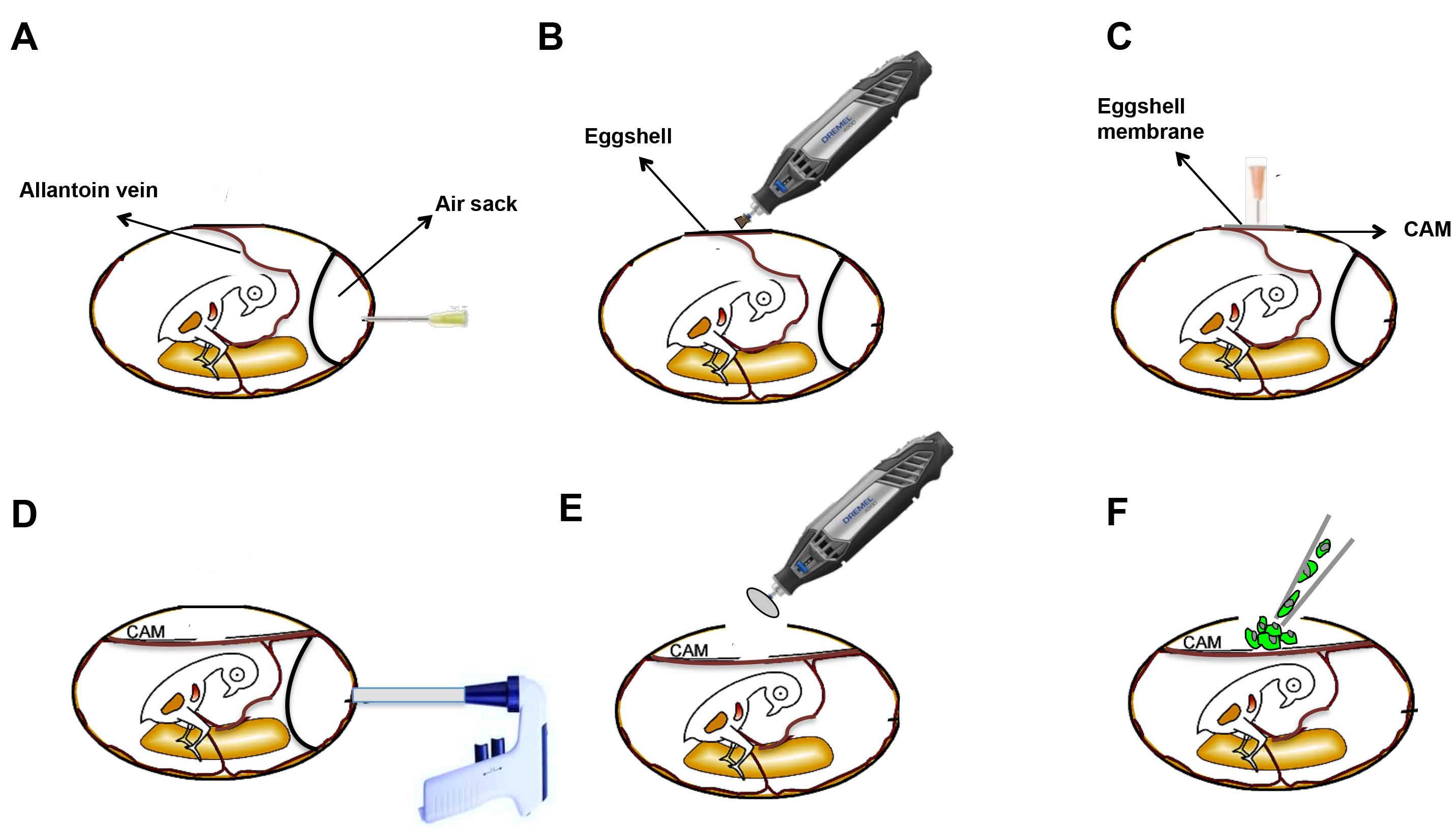 Figure 1. Schematic Illustration of the CAM angiogenesis assay.
Materials and Equipment
Fertilized E6 chicken embryos
0.1% Benzalkonium bromide
Methanol and acetone (1:1 in volume)
Filter-paper disk;
Parafilm (packing film)
Normal saline
Incubator
Glass slide
Ophthalmic forceps
Assay Procedure
- Fertilized E6 chicken embryos (48 ± 5 g) are cleaned with 0.1% benzalkonium bromide and pre-incubated at 37.5°C in 85% humidity for 2 days.
- Egg morphology appears like a meta-ellipse, with a relatively larger side and a smaller one, and the air sac is usually located on the larger side right behind the shell. After disinfection of the shell center outside the air sac with 0.1% benzalkonium bromide, a hole highlighted with marker pen is buffed and drilled gently over the air sac with a nipper not to break the shell, and the vascular zone is easy to be identified on the CAM.
- Two drops of normal saline are then added to moisten the inner shell membrane adjacent to the CAM so that the membrane is easy to be separated from CAM.
- After being clamped and raised by ophthalmic forceps, the membrane and the CAM separated unforcedly, and then a 1 x 1 cm window on the membrane is sectioned to expose the vascular zone.
- A 5 mm x 5 mm sterilized filter-paper disks, which are used as a carrier for being directly loaded with indicated concentrations of chemicals or virus, are then directly applied and adhere to the vascular zone with right density of vascular.
- Upon sealing the openings with sterile flexible packing film, the eggs are further incubated for indicated periods.
- Finally, a mix of methanol and acetone (1:1 in volume) is directly added to immerse and fix the blood vessels of the experiment zone.
- After being clamped and raised by ophthalmic forceps, the CAM is easy to be separated from the embryo, and it is cut and spread on glass slide, and the blood vessels are viewed, photographed and quantified by counting the number of blood vessel branch points.
Troubleshooting
- False-positive responses
One of the caveats of the CAM assay is the high number of spontaneous positive responses obtained. In addition, false-positive responses can also be caused by the presence of salt crystals in the sample or pieces of eggshell that become lodged inside the egg. Although some of these issues can be addressed, often they are beyond the control of the investigator. - Another serious problem that can reduce embryo viability and/or the number of samples is contamination.
This problem can be reduced by following more stringent sterile techniques including the use of sterile equipment, careful swiping of the eggs with betadine or 70% alcohol before opening, working in a sterile hood in extreme cases, or maintaining a low incubator humidity.
References
- Ribatti D, et al.; The chick embryo chorioallantoic membrane as a model for in vivo research on angiogenesis. Int J Dev Biol, 1996, 40: 1189-1197.
- Ponce M L, et al.; The chick chorioallantoic membrane as an in vivo angiogenesis model. Current Protocols in Cell Biology, 2003, 18(1): 19.5.1-19.5.6.
- West D C, et al.; Angiogenesis assays using chick chorioallantoic membrane. Methods Mol Med, 2001, 46: 107-129.
Cell Services:
Cell Line Testing and Assays
